43 lungs pictures with labels
Lung diagram | Lungs image | Simple lungs diagram | Lung anatomy ... Lung anatomy diagram or Simple lungs diagram with label are also mentioned below. Pharmacy Images 372 followers More information You can clearly understand by observing the Lung diagram in this post.We are providing simple lungs diagram for quick drawing the diagram. You can also download lungs image that are given in the post. CT chest lung window axial - labeling questions - Radiopaedia Normal CT chest lung windows (with labels) Annotated image Axial lung window The labeled structures are (excluding the correct side): cervical portion of trachea apical segment of RUL apicoposterior segment of LUL thoracic portion of trachea thoracic portion of esophagus left oblique (major) fissure apical segmental bronchus of RUL
Respiratory System With Labels Pics stock illustrations Browse 2,431 respiratory system with labels pics stock illustrations and vector graphics available royalty-free, or start a new search to explore more great stock images and vector art.

Lungs pictures with labels
Histology Illustrations - Bronchi and lungs (labels) - illustration Bronchi and lungs (labels) - illustration From ; funded by the U.S. National Cancer Institute's Surveillance,Epidemiology and End Results (SEER) Program, via contract number N01-CN-67006, with Emory University, Atlanta SEER Cancer Registry, Atlanta, Georgia, U.S.A. 3D model Normal heart and lungs - AnatomyTOOL This is a model of the normal heart and lungs. It shows the left and right ventricles, the left and right atria, the aorta and vena cava superior and inferior and the pulmonary trunk and pulmonary arteries and the pulmonary veins. Also both lungs are shown. Label The Lungs Photos and Premium High Res Pictures - Getty Images Find Label The Lungs stock photos and editorial news pictures from Getty Images. Select from premium Label The Lungs of the highest quality. CREATIVE. ... 19 test tube blood with label result on the tube with lung x ray background, coronavirus disease (covid-19) concept. - label the lungs stock pictures, royalty-free photos & images.
Lungs pictures with labels. drawing of lungs with labels File:Diagram of the human heart (cropped).svg - Wikimedia Commons we have 9 Pictures about File:Diagram of the human heart (cropped).svg - Wikimedia Commons like Diagram of Air Tubes in the Lungs | ClipArt ETC, Respiratory System With Label Drawing at GetDrawings.com | Free for and also Simple Pavement Epithelium Cells | ClipArt ETC. Lungs: Definition, Location, Anatomy, Function, Diagram, Diseases Where are the Lungs Located. The lungs are located a little toward the posterior part of the human body, just below the collarbone, extending down to the diaphragm, the muscular partition that separates the chest and abdominal cavities.The left and right lungs are situated on the two sides of the body with the heart, another vital organ in the thoracic cavity, located a little in front of, and ... Labeled diagram of the lungs/respiratory system. - SERC Labeled diagram of the lungs/respiratory system. Image 37789 is a 1125 by 1408 pixel PNG. Uploaded: Jan10 14. Last Modified: 2014-01-10 12:15:34. Permanent URL: . The file is referred to in 1 page. Airborne Microbes. Lungs: Anatomy, Function, and Treatment - Verywell Health SCIEPRO/SCIENCE PHOTO LIBRARY Anatomy Structure . ... Lung tissue diseases like pulmonary fibrosis and sarcoidosis. There are 30,000 to 40,000 new cases of pulmonary fibrosis diagnosed in the U.S. each year, affecting 100,000 people in total. Sarcoidosis is considered a rare disease, affecting fewer than 200,000 in the U.S. ...
Lungs Picture Image on MedicineNet.com Picture of Lungs The lungs are a pair of spongy, air-filled organs located on either side of the chest (thorax). The trachea (windpipe) conducts inhaled air into the lungs through its tubular branches, called bronchi. The bronchi then divide into smaller and smaller branches ( bronchioles ), finally becoming microscopic. Normal CT chest lung on axial images with labels - IMAIOS Normal thoracic CT on axial images. We created an anatomy atlas of the chest and the mediastinum which is an interactive tool for studying the cross-sectional anatomy of the normal thorax based on an enhanced multidetector computed tomography with helical angiography of the thorax (axial plane). Anatomical structures are visible as interactive ... Human Lung Photos and Premium High Res Pictures - Getty Images human lung organ 18,343 Human Lung Premium High Res Photos Browse 18,343 human lung stock photos and images available, or search for human lung anatomy or human lung illustration to find more great stock photos and pictures. Related searches: human lung anatomy human lung illustration healthy human lung human lung alveoli human lung cells of 100 Labeled Diagram of the Human Lungs - Bodytomy Given below is a labeled diagram of the human lungs followed by a brief account of the different parts of the lungs and their functions. Each lung is enclosed inside a sac called pleura, which is a double-membrane structure formed by a smooth membrane called serous membrane.
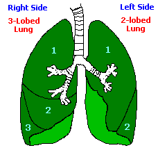
Picture Illustration of Anatomical Structures - Lungs Picture of Lungs. The lungs are organs used for breathing located on either side of the chest. The lungs fill with air, oxygenate the blood, and dispose of carbon dioxide. Lungs are comprised of many different structures. The image on this page depicts the trachea, bronchi, and the several lobes of the left and right lungs. Label Lungs Diagram Printout - Enchanted Learning right inferior lobe: the bottom lobe of the lung on the right side of the body. right middle lobe: the middle lobe of the lung on the right side of the body. right superior lobe: the top lobe of the lung on the right side of the body. 298,135 Lungs Images, Stock Photos & Vectors | Shutterstock Find Lungs stock images in HD and millions of other royalty-free stock photos, illustrations and vectors in the Shutterstock collection. Thousands of new, high-quality pictures added every day. CT chest lung window sagittal - labeling questions - Radiopaedia Normal CT chest lung windows (with labels) Annotated image. Loading Image 1. Annotated image. Sagittal lung window. The labeled structures are (excluding the correct side): lateral costophrenic recess. lateral basal segment of LLL. left oblique (major) fissure.

White ramus communicans - wikidoc
Label the lungs Vector Art Stock Images | Depositphotos Discover 198 Label the lungs vectors in the Depositphotos collection Premium vector graphics scalable to any size. Feel free to use images in art designs!

Lecture 28 - Lungs flashcards | Quizlet
Lung Anatomy, Function, and Diagrams - Healthline The lungs begin at the bottom of your trachea (windpipe). The trachea is a tube that carries the air in and out of your lungs. Each lung has a tube called a bronchus that connects to the trachea....

Lung Labeling Quiz
Diagram Of The Respiratory System With Labels Stock Photos, Pictures ... Lungs with Alveoli Labeled CG image of woman's chest area showing both lungs in isolation, with magnified view of alveoli air sacs labeled on faded flesh tone and white. diagram of the respiratory system with labels stock pictures, royalty-free photos & images
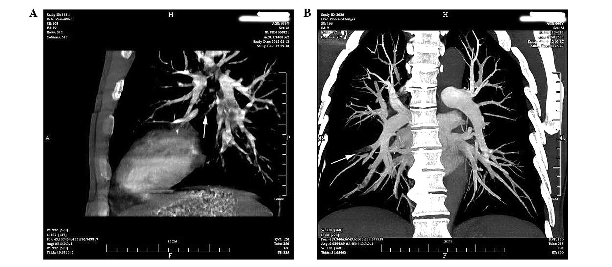
Application of 128‑slice spiral CT combination scanning in the diagnosis of embolisms in ...
Label The Lungs Photos and Premium High Res Pictures - Getty Images lungs. flat icons on buttons in different colors - label the lungs stock illustrations close-up hands wearing gloves holding covid 19 test tube blood with label result on the tube with lung x ray background, coronavirus disease (covid-19) concept. - label the lungs stock pictures, royalty-free photos & images

Respitory system
Lung Histology - Bronchi and lungs (labels) - illustration Bronchi and lungs (labels) - illustration From ; funded by the U.S. National Cancer Institute's Surveillance,Epidemiology and End Results (SEER) Program, via contract number N01-CN-67006, with Emory University, Atlanta SEER Cancer Registry, Atlanta, Georgia, U.S.A.

Lung Stuff - Home
Label The Lungs Photos and Premium High Res Pictures - Getty Images 187 Label The Lungs Premium High Res Photos Browse 187 label the lungs stock photos and images available or start a new search to explore more stock photos and images. of 4 NEXT

Lungs Quiz
Human Lung Stock Photos, Pictures & Royalty-Free Images - iStock Browse 63,269 human lung stock photos and images available, or search for human lung anatomy or human lung illustration to find more great stock photos and pictures. 3D illustration of Lungs, medical concept. 3D illustration of Lungs - Part of Human Organic.

add lungs
These fee printable human lung worksheets include coloring pages, label ... Human Lungs Worksheets These free printable human lung worksheets include coloring pages, label worksheets, notebooking pages, and more! Incorporate them into your science day or unit study for excellent anatomy resources. C Clare 8 followers More information

Everything you need to know about the lungs in just 5 minutes - YouTube
Lungs (Human Anatomy): Picture, Function, Definition, Conditions The lungs are a pair of spongy, air-filled organs located on either side of the chest (thorax). The trachea (windpipe) conducts inhaled air into the lungs through its tubular branches, called...

Lungs and Trachea | ClipArt ETC
Lung Diagram Labelling Activity | Primary Resources | Twinkl This handy Lung Labelling Worksheet gives your children the opportunity to show how much they've learned about the human lung system. The beautifully hand-drawn illustration shows a lung diagram, labelled with blank spaces where learners can fill in its different components. Encourage your students to work independently and label the parts of the lungs they can see. This teaching resource also ...
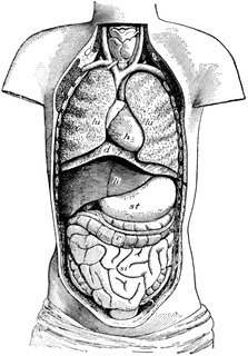
Ventral Cavity of the Body | ClipArt ETC
Label The Lungs Photos and Premium High Res Pictures - Getty Images Find Label The Lungs stock photos and editorial news pictures from Getty Images. Select from premium Label The Lungs of the highest quality. CREATIVE. ... 19 test tube blood with label result on the tube with lung x ray background, coronavirus disease (covid-19) concept. - label the lungs stock pictures, royalty-free photos & images.

Circulation and respiration | Circulatory and respiratory systems | Siyavula
3D model Normal heart and lungs - AnatomyTOOL This is a model of the normal heart and lungs. It shows the left and right ventricles, the left and right atria, the aorta and vena cava superior and inferior and the pulmonary trunk and pulmonary arteries and the pulmonary veins. Also both lungs are shown.

The lungs - YouTube
Histology Illustrations - Bronchi and lungs (labels) - illustration Bronchi and lungs (labels) - illustration From ; funded by the U.S. National Cancer Institute's Surveillance,Epidemiology and End Results (SEER) Program, via contract number N01-CN-67006, with Emory University, Atlanta SEER Cancer Registry, Atlanta, Georgia, U.S.A.

Lungs, Health and Search on Pinterest
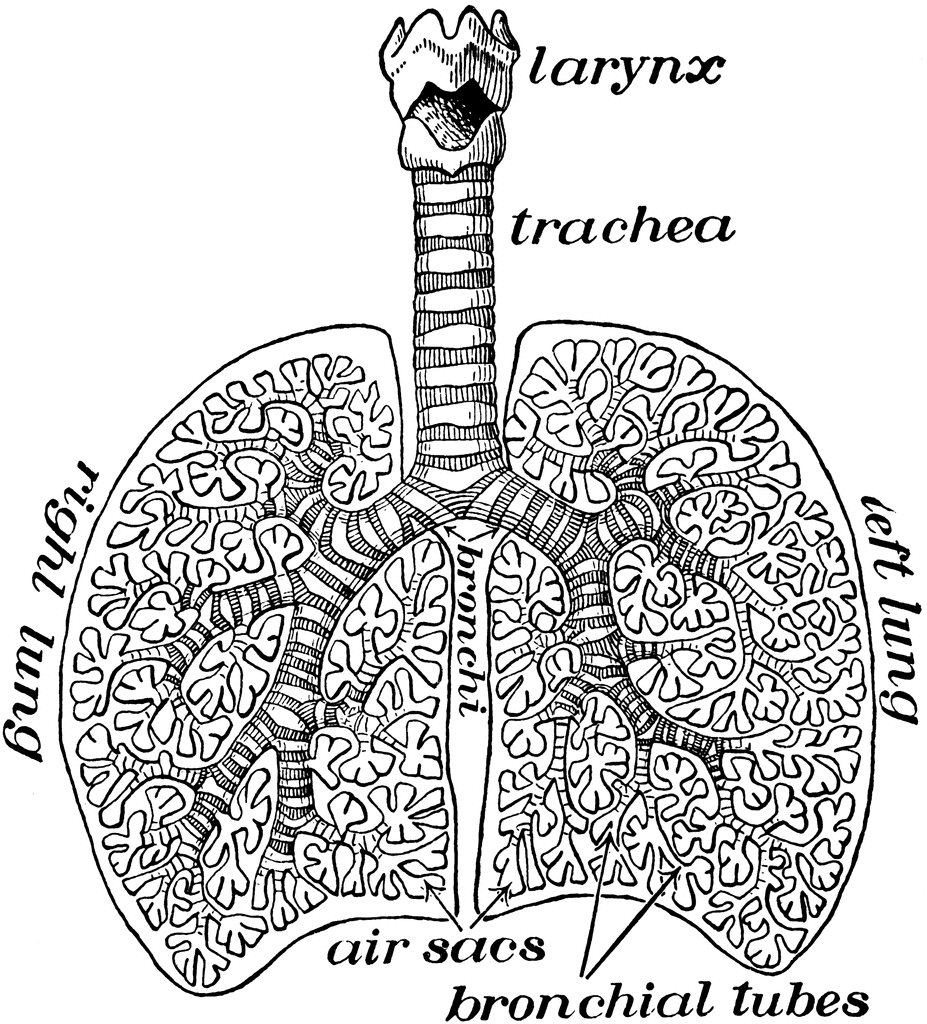
Diagram of Air Tubes in the Lungs | ClipArt ETC

Lungs - YouTube

Cells, major tissue types , Epithelial Cells Flashcards | Easy Notecards
Threesology Research Journal
Post a Comment for "43 lungs pictures with labels"